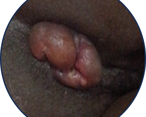
Hemorrhoids are tortuous dilatation and elongation of the venous plexus and overlying cushion in the wall of the anus. Hemorrhoids often present with bright red bleed, edema and prolapse of the anal cushions.
Pathology
Hemorrhoids are diagnosed clinically when the hemorrhoidal cushions located in the walls of the anus begin to give symptoms. There are the major and the minor cushions; the major cushions are located at specific locations (3, 7 & 11 o’clock) whereas the minor ones are in between the locations of the major ones.
Structure of the Anal Cushions
The anal cushion is part of the normal anatomical and functional structures of the anal canal.
The anal cushions comprise of 3 main structures
(1) arteriovenous communcations called sinusoids,
(2) overlying anal mucosa and
(3) suspensory ligaments called ligaments of Treitz
The sinusoids are formed by the branches of the superior hemorrhoidal artery, which supply blood to the cushions. The anal mucosa is a stratified squamous epithelium that covers the cushions and forms the cutaneous zone of the anal canal. The ligaments of Treitz are thin bands of connective tissue that attach the cushions to the surrounding muscles and skin.
The function of the anal Cushions
- Sensing content of rectum: differentiating the sensation flatus and feaces
- Contribute to anal continence. Responsible for 15-20% of normal resting pressure,
The mucosa is composed of 2 layers; the transitional epithelium and subepithelial connective tissue
The suspensory ligament which supports the cushion has 2 components; the subepithelial muscle and the parks ligament attaching to the internal sphincter. Together the 2 components of the suspensory ligament are referred to as the Treitz muscle
The hemorrhoidal arteries (superior, middle and inferior ) are the arterial inflow to the sinusoids and the hemorrhoidal veins (superior, middle and inferior) are the venous outflow from the sinusoids
Generally, Symptoms of hemorrhoid arise from enlargement, stretching and prolapse of these hemorrhoidal cushions structures and the theories of initiation or etiology and progression or evolution of hemorrhoids revolve around changes in the anal cushions.
Pathogenesis
Several theories have been proposed in the pathogeneiss of hemorrhoids. In this discussion we will describe 2 groups of theories
Theory of ethiolgy and theory of evolution; the theory of etiology are theories that attempt to describe the initiating factors for hemorrhoids appears or direct identifiable cause.
The theory of evolution are those describing the structural change underpining the formation of hemorrhoids
Four theories of Etiology /Pathophysiology Events
- Morgan’s theory: This theory proposes that the hydrostatic pressure of erect posture causes engorgement in the hemorrhoidal sinusoids due to the absence of valves in the inferior vena cava (IVC) and portal venous system.
- Durel’s theory: This theory suggests that sitting to defecate, straining at defecation, and prolonged sitting on a toilet bowl impair venous return from the pelvis and cause increased pelvic pressure and tourniquet effect on the hemorrhoidal veins.
- Park’s postulate: This postulate states that impacted feces above the dentate line obstructing hemorrhoidal venous return cause engorgement and prolapse.
- Alexander William’s Theory: This theory describes a vicious circle of increased anal canal pressure, hemorrhoid prolapse caught by sphincter with further congestion.
These theories are based on empirical observations and clinical experiences, but they are not supported by conclusive evidence or scientific principles. They do not explain how hemorrhoids are formed or how they can be prevented or treated
Four theories of Evolution or formation
- Sliding mucosa Cushion Theory: This theory proposes that the hemorrhoidal cushions slide down when the suspensory structures weaken and stretch. This is the most widely accepted theory. Inflammatory mediators, such as proteases and matrix metalloproteinases, are thought to weaken the structures and degrade the extracellular proteins, such as elastin, fibronectin, and collagen, in the hemorrhoidal tissue.
- Rectal Varicosity from portal hypertension (John Hunter’s): This theory suggests that hemorrhoids are caused by varicose veins in the rectum due to portal hypertension. This theory has been debunked because varicosities and hemorrhoids are described as different entities in the anus.
- Rectal mucosal redundancy theory: This theory states that hemorrhoids are formed by spontaneous elongation of the cushions, mucosa, and suspensory ligaments. This theory is based on the observation that some patients with hemorrhoids have redundant rectal mucosa.
- Vascular hyperplasia and abnormality (Virchows and Allingham liken hemorrhoid to hemangioma): This theory describes hemorrhoids as a type of hemangioma, a benign tumor of blood vessels. This theory is based on the evidence of angioproliferation and neovascularity in the hemorrhoidal tissue, due to increased levels of transforming growth factor-beta and vascular endothelial growth factor. This theory also suggests that there is dysregulation of venous tone in the hemorrhoidal veins.
These theories are based on empirical observations and clinical experiences, but they do not explain the exact mechanisms or factors that lead to the formation of hemorrhoids. They also do not account for the genetic, environmental, and lifestyle influences that may contribute to the development of hemorrhoids. Therefore, the etiology and pathophysiology of hemorrhoids remain unclear and controversial.
Classification of hemorrhoids (7 ways to classify)
The classification of hemorrhoids can be based on different criteria, such as:
- The cause: idiopathic or secondary to a specific event or situation, such as portal hypertension, pregnancy, or chronic constipation1.
- The location around the anorectal ring: primary locations corresponding to the termination of the superior hemorrhoidal arteries (3, 7, and 11 o’clock positions) and secondary locations in between the primary ones.
- The origin in relation to the dentate line: internal (above the dentate line) or external (below the dentate line). Internal hemorrhoids are covered by mucosa, while external hemorrhoids are covered by skin.
- The degree of prolapse: this is a functional or clinical classification used for internal hemorrhoids only. It ranges from grade 1 (no prolapse) to grade 4 (irreducible prolapse).
- The morphology: pedunculated (with a stalk) or sessile (without a stalk).
- The histology: fibrous (with thickened connective tissue) or vascular (with dilated blood vessels).
- The symptoms: asymptomatic or symptomatic (with bleeding, pain, itching, or discharge).
Practical Grading of Internal hemorrhoids
Practical grading of internal hemorrhoids is helpful when selecting treatment
Based on the degree of prolapse (Originally Goligher’s/ Banov et al. Grading of internal hemorrhoids 1985)
1st degree: Bleeding with no external prolapse during defecation
2nd degree: prolapses during but reduces spontaneously
3rd degree: Prolapse needing to be digitally returned by the patient
4th degree Stays out permanently with or without defecation.
Some examples of treatment options for different grades of internal hemorrhoids are:
- Grade 1: Conservative measures, such as dietary modification, increased fluid intake, stool softeners, topical agents, and sitz baths.
- Grade 2: Office-based procedures, such as rubber band ligation, sclerotherapy, infrared coagulation, or bipolar diathermy.
- Grade 3: Surgical procedures, such as hemorrhoidectomy, stapled hemorrhoidopexy, or hemorrhoidal artery ligation.
Grade 4: Surgical procedures, such as hemorrhoidectomy or stapled hemorrhoidopexy, with possible additional interventions for complications, such as thrombosis, strangulation, or infection
